How Periapical X-Rays Can Help Dental Extraction?
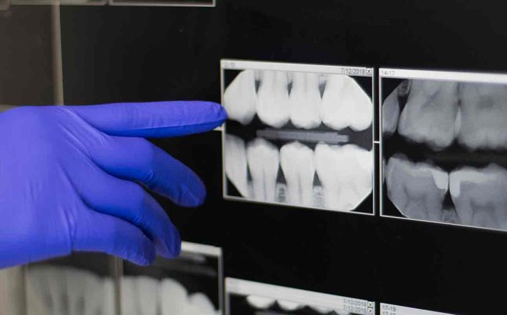
You might be curious about how the dentist evaluates the extraction’s success when you undergo an operation like that. The periapical x ray is a crucial tool in this evaluation. This article will explain how periapical X-rays are used to assess the effectiveness of tooth extraction treatments.
Examining The Periapical X-Ray
Locating The Problem
Finding the precise issue is one of the main goals of a periapical X-ray prior to extraction. It aids the dentist in locating problems like cavities, infections, or tooth damage. Planning a successful extraction requires having a clear understanding of the problem’s location.
Assessing Root Structure
Your teeth’s roots are not visible to the naked eye because they are covered by the gum line. However, periapical X-rays show the root structure. This information is essential because it enables the dentist to identify any anomalies, such as curved or hooked roots. The dentist can take the help of eruption chart teeth and then make appropriate plans for the extraction procedure.
Evaluating Bone Density
For a tooth extraction to be successful, the bone surrounding the tooth must be healthy and dense. An image of the bone structure is produced by a periapical X-ray, which shows whether there is sufficient healthy bone to sustain the extraction procedure. To ensure a successful extraction, further operations could be required if the bone is too fragile or injured.
During The Extraction
Guidance During The Procedure
The periapical X-ray continues to be important while you are in the dental extraction near me. The X-ray can be used by the dentist to confirm that the entire tooth, including all of its roots, is being extracted. This step is essential to stop any leftover pieces from complicating the situation.
Monitoring The Progress
The dentist may take more periapical X-rays while the extraction goes on to evaluate how the tooth is being removed. These X-rays offer immediate feedback, assisting the dentist in making the required corrections to guarantee a painless and effective extraction.
Post-Extraction Assessment
Ensuring Complete Removal
A second periapical X-ray is taken by the dentist once the extraction is finished. The entire tooth, including all roots and pieces, has been successfully extracted, according to this post-extraction X-ray. The danger of infection or other problems is decreased by ensuring thorough clearance.
Checking For Complications
Complications can occasionally arise even after a successful extraction. Periapical X-rays are helpful for post-extraction evaluation because they let the dentist look for any problems, including infection, damage to the teeth next to the extracted tooth, or bone fractures. For prompt treatment, it is essential to identify such issues early.
Tracking Healing Progress
Your body begins the healing process while you recuperate from the extraction. During follow-up visits, periapical X-rays may be done to monitor the course of the healing. These X-rays demonstrate how the bone is reclaiming the tooth socket after it was pulled. Monitoring the healing process makes sure that everything is going according to plan.
Conclusion
In conclusion, periapical X-rays are a crucial tool for emergency tooth extractions near me for determining whether tooth extraction treatments were successful. These X-rays are useful for pre-extraction planning, during the extraction, and for post-extraction evaluation. They offer a thorough view of the tooth, its roots, the surrounding bone, and any possible problems. Contact wisdom tooth removal near me for any complications.
- SHARES


